When someone dies with suspected lung disease, families want clear answers. A lung-only autopsy gives focused answers about lung problems. It does not require a full-body examination. Understanding what doctors find helps families make informed decisions.
What Is a Lung Only Autopsy?
A lung only autopsy is a focused exam. It looks only at the lungs and chest area.
Unlike regular autopsies, this procedure checks the lungs and chest wall. It also checks pleural surfaces and nearby structures. The goal is to identify disease or exposure problems.
The medical examiner performs these exams. Sometimes the department of pathology does them. These exams happen when specific questions exist about lung disease. They also happen when there is a history of asbestos exposure.
The process includes removing the lungs. It includes detailed visual checks. It also includes looking at a tissue sample under a microscope. Many families choose this option when they need answers about lung conditions.
The Lung Autopsy Process
Visual Examination
The lung pathology autopsy process starts with visual examination. Doctors call this gross examination lung autopsy. During this phase, they observe features visible to the naked eye.
They check lung size. They check weight, color, and texture. The chest wall and pleural surfaces get careful attention. Problems here often point to exposure-related disease.
Doctors document pleural plaques. They note thickening areas, masses, and any fluid buildup.
They examine airways, blood vessels, and lymph nodes. Each observation gives clues about disease. It also gives clues about exposure history. This visual exam sets the stage for microscopic work.
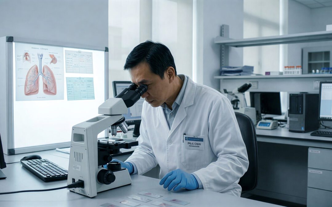
Microscopic Examination
After the visual exam, pathologists select tissue samples. These come from different lung areas.
Someone processes these samples. They cut the samples into thin sections for microscope viewing. Under the microscope, cell details emerge. You cannot see these details otherwise.
Microscopic lung autopsy findings show many things. They show inflammation patterns. They show scarring and cell changes. They show foreign materials like asbestos fibers.
Doctors identify specific cell types. They look for signs of occupational exposure.
This detailed examination often gives definitive answers. It explains diseases that were unclear during life.
Pleural Plaques
Pleural plaques are common asbestos related lung pathology findings. People raised these areas. They appear white or gray.
They form on the pleural surface. They typically appear on the chest wall or diaphragm. They consist of dense tissue. They show past asbestos exposure.
During a lung autopsy for asbestos exposure, doctors document where plaques appear. They note how large they are.
Plaques do not cause chest pain. They do not cause symptoms. They do not turn into cancer.
However, they prove someone breathed in asbestos fibers. This matters for family members. They may have shared similar environments.
Under the microscope, pleural plaques show a specific pattern. Doctors can tell these apart from other types of thickening. They have distinct features.
Recording pleural plaques pathology findings helps families. It helps them understand exposure history. It supports legal claims.
Understanding Pleural Rind
The term “pleural rind” describes a different problem. Different than separate plaques.
Pleural rind pathology meaning refers to a continuous, thick layer. This layer wraps around the lung.
It can result from inflammation. It can result from infection. It can also result from asbestos exposure.
In asbestos cases, thick pleura may form a restrictive layer. This layer limits lung expansion.
Doctors measure this layer’s thickness. They check whether it covers the visceral pleura. They check if it covers the chest wall. Unlike separate plaques, extensive rind formation causes serious breathing problems during life.
Microscope exam reveals what the thickened pleura contains. In asbestos cases, doctors often find scarring. They find it mixed with inflammation.
Finding asbestos fibers in the tissue confirms exposure as the cause. This distinction matters for legal purposes. It matters for compensation purposes.
Pleural Thickening vs Lung Cancer
One critical part of lung autopsy pathology explained involves telling benign problems from cancer. Pleural thickening vs lung cancer represents an important comparison. This requires careful review.
Both conditions can show similar symptoms. Both can show similar chest x rays.
This makes postmortem exam valuable. It provides definite diagnosis.
Benign pleural thickening appears as smooth, fibrous layers. It does not show destructive growth.
The tissue shows organized structure. It has minimal cell activity.
In contrast, cancer shows irregular growth patterns. It shows invasion into lung tissue. It shows abnormal cells.
During lung cancer differential diagnosis pathology, examiners look for specific patterns. They look for abnormal cells. They look for invasion signs. They may use special stains. These confirm cell type.
The postmortem mesothelioma diagnosis gives definitive answers. It helps when doctors were unsure during life.
Asbestos Related Diseases
Asbestos exposure creates several distinct patterns. These go beyond pleural changes.
Pathologists look for lung scarring. This particularly appears in lower lobes. This marks asbestosis.
They identify asbestos bodies. These are fibers coated with iron. They appear as golden-brown structures under the microscope.
The pattern and amount of scarring help doctors. They use it to grade asbestosis severity.
They check whether scarring extends into air spaces. This is a pattern specific to this occupational disease. Special iron stains confirm fiber burden.
You can count asbestos fibers. This measures exposure.
This specialized examination gives objective evidence. It shows exposure levels.
When combined with medical history and autopsy findings, fiber analysis strengthens the connection. It connects exposure and disease.
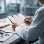
The Autopsy Report
After completing visual and microscope exams, pathologists write their findings. They write a comprehensive report. A lung autopsy report explained includes several key sections. These document the exam and conclusions.
The autopsy report starts with medical history. It includes the reason for examination. It describes visual findings in detail. This includes measurements. It includes visible problems.
The microscope section details cell-level observations. These come from each sampled area. Doctors include descriptions of special stains performed.
The final diagnosis section summarizes key findings. It gives interpretations.
This may include statements about asbestos exposure. It may include disease type. It may include potential causes of death.
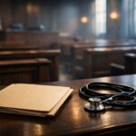
The report serves as a permanent medical document. It serves as a legal document for families.
Why Families Choose These Services
When searching for lung autopsy services near me, families want answers. They have specific questions.
Did work exposure cause the disease? What exactly was wrong? Could earlier diagnosis have helped?
These questions drive the decision. They lead to pursuing limited examination.
Many families choose lung-only exam. It gives focused answers. It does not have the emotional burden of full autopsy.
The procedure respects the body. It gets necessary information.
Results support legal claims. They inform family members about exposure risks. They provide closure through definite diagnosis.
Specialized services understand the sensitive nature of these exams. They work with families. They explain the process. They explain the timeline. They explain what information the exam can provide.
The lung only autopsy offers a respectful approach. It gets critical medical information.
What Doctors Look For
How pathologists examine lungs follows a step-by-step protocol. They start by observing external surfaces. They note any adhesions. They note plaques or masses.
After documenting external features, they make cuts. These cuts examine internal structures.
Airways get careful inspection. Doctors check for blockage. They check for inflammation or tumors.
Doctors check blood vessels for clots. Someone checks them for other problems. Doctors assess the lung tissue for nodules. Assessed for scarring or damage.
We examine each section separately. This identifies regional differences.
Taking samples for microscopy follows a standard protocol. Pathologists take sections from each area. They take them from any visible problems. They take them from normal-appearing areas for comparison.
This thorough sampling ensures that we find diseases. It finds both focal and widespread diseases.
Other Lung Diseases Found
Asbestos-related problems are common reasons for lung-only autopsy. However, doctors identify many other conditions during these exams.
Doctors can diagnose lung diseases. Doctors can diagnose infections. Doctors can diagnose emphysema. Doctors can diagnose various cancers through postmortem examination.
Work-related lung diseases beyond asbestosis include silicosis. They include coal worker’s disease. Each has characteristic features. Doctors recognize these features.
The exam may reveal unexpected findings. These explain puzzling symptoms. They explain breathing decline.
Sometimes doctors find conditions like pulmonary edema. These contributed to death.
The medical center documents all significant findings. The department of pathology does this in autopsy cases. This provides a complete picture of lung problems.
For those who want to find articles about specific conditions, the autopsy report provides detailed documentation. It helps researchers and family members understand the full scope of disease.
Frequently Asked Questions
What do doctors examine during a lung-only autopsy?
Doctors examine both lungs. They examine the pleural membranes. They examine major airways, blood vessels, and lymph nodes.
The exam includes visual inspection. It includes microscopic examination of tissue samples from multiple lung regions.
How long does the process take?
The physical exam takes several hours. The complete process usually requires two to four weeks. This includes microscope examining. It includes report preparation.
Can this autopsy diagnose mesothelioma?
Yes. Pathologists can definitely diagnose mesothelioma through lung-only autopsy. The exam reveals characteristic growth patterns. It reveals cellular features specific to mesothelioma.
What do pleural plaques mean?
Pleural plaques provide clear evidence. They show past asbestos exposure. They develop years after exposure. They prove asbestos fibers reached the pleural space.
How do doctors tell pleural thickening from lung cancer?
Doctors use multiple criteria for differential diagnosis. Benign thickening shows organized tissue. It does not show invasion.
Cancer shows destructive growth. It shows abnormal cells.
CT scan findings during life may have suggested problems. However, microscope exam provides definite answers.
Will this affect funeral plans?
Lung-only autopsy minimizes visible changes. It allows normal funeral arrangements. This includes open casket services. The procedure focuses on the chest area only.
What will the report include?
The report includes detailed descriptions. It describes visual findings. It describes microscope observations.
It includes results of special studies. It includes a final diagnosis. It documents all significant problems. It provides interpretations about disease processes.
Can these findings support legal claims?
Yes. Lung autopsy findings frequently support legal claims. These relate to occupational exposure. The detailed report provides objective scientific evidence. This carries significant weight in legal proceedings.
How accurate is this compared to clinical diagnosis?
Lung-only autopsy provides the gold standard for diagnosis. Studies show autopsy reveals unexpected findings. It corrects diagnoses in many cases.
Direct tissue examination provides certainty. Chest x rays cannot match this. CT scan cannot match this certainty.
Can family members get copies of the report?
Yes. Family members receive copies of the complete report. The report becomes part of medical records. Doctors can share it. The family can share it with attorneys as they direct.