The loss of a loved one can be confusing and painful, and many families are left looking for answers. A brain-only autopsy offers an intricate exploration of possible neurological factors contributing to the death. This extensively expanded guide delves into the complete process, from initial preparation to comprehensive analysis and reporting. Each section builds on the previous one to provide families and readers with a detailed, informed perspective. By expanding the scope and depth of information, this document serves as a valuable resource for those seeking clarity.
Comprehensive and Detailed Brain Examination Process
The process begins with family consent and an in-depth review of the decedent’s medical history. The forensic pathologist carefully plans the procedure, incorporating any family concerns and questions.
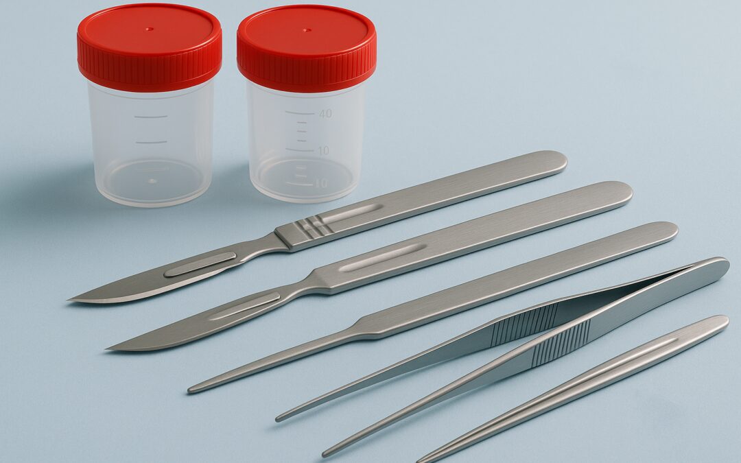
The brain is removed during the autopsy with great precision, maintaining respect and dignity. Additional care is taken to minimize cosmetic disruptions, which can be important for viewing arrangements.
Advanced tracking systems and strict identification protocols are used to ensure accuracy and integrity of the specimen throughout. Additional security measures, including the use of tamper-evident seals, may be implemented to further protect the specimen during analysis.
In certain cases, coordination with funeral homes or religious officials may be necessary to align the procedure with family customs.
Extended Brain Retrieval and Examination Procedures
Once retrieved, the brain is examined externally to detect trauma, infections, or signs of neurodegenerative disease.
Advanced imaging techniques like MRI, CT scans, and 3D digital modeling often complement the physical examination, offering a more comprehensive view of brain structures.
The brain is sliced into extremely thin sections and preserved in formalin to maintain tissue quality.
Each step is digitally documented, and supplementary imaging captures the specimen’s state at various stages, forming a permanent and comprehensive record.
Additional procedures, such as advanced colorimetric staining, may be employed to enhance visualization of specific structures or abnormalities.
In complex cases, additional consultations with forensic teams may guide the examination plan.
Advanced Brain Tissue Analysis and Processing
In this expanded phase, forensic pathologists employ advanced staining techniques and immunohistochemistry to highlight disease markers and structural abnormalities.
High-resolution digital microscopes and AI-assisted imaging systems enhance detection accuracy.
Specialized analysis identifies subtle cellular changes linked to Alzheimer’s, Parkinson’s, and other conditions.
All tissue samples are cataloged, labeled, and stored under stringent conditions, ensuring they remain available for future reference, legal needs, or research.
Advanced computational tools, such as digital pathology software and machine learning algorithms, help analyze complex tissue data, elevating diagnostic precision.
For example, machine learning can identify subtle cellular patterns in brain tissue that indicate early stages of diseases like Alzheimer’s, while digital pathology platforms enable automated quantification of tissue markers, improving consistency and accuracy.
Additional genetic sequencing techniques may be applied to detect hereditary conditions.
Pathologists may also consult with genetic counselors to inform families of potential hereditary implications.
Deepened Forensic Neuropathology Correlation
Forensic neuropathologists meticulously correlate microscopic findings with the individual’s health records, symptoms, and imaging history.
This expanded correlation can reveal silent strokes, early-stage neurodegenerative changes, or previously undiagnosed infections.
Insights from this phase can inform family health considerations, including hereditary risks, and contribute to ongoing public health research.
The findings not only serve individual families but also enhance collective medical understanding of brain diseases.
Newer techniques like proteomic analysis may provide even deeper insights into complex neurological conditions.
Further, in some cases, cross-referencing with other postmortem investigations can offer a more holistic understanding of the cause of death.
Expanded Diagnostic Reporting Process
Upon conclusion of the examination, the forensic pathologist prepares a detailed, multi-layered report.
This report incorporates macroscopic and microscopic findings, digital images, annotated diagrams, and comprehensive interpretations.
Preliminary results may be provided within days, while the complete report can take several weeks.
The final document includes detailed explanations, photographic evidence, and context about how the findings relate to the individual’s medical history.
Families are encouraged to consult with the forensic expert to fully understand the report.
Support services, including counseling or additional medical consultations, may also be offered to help families interpret the findings and plan for the future.
In some cases, families may request second opinions or additional reviews by independent specialists.
Enhanced Postmortem Brain Analysis Steps
- Consent and Preliminary Review: Families provide informed consent, and the pathologist conducts a detailed review of medical and clinical records.
- Brain Removal: The brain is removed with precision, preserving the body’s cosmetic appearance.
- Preservation and Documentation: The brain is fixed in formalin, sectioned, and digitally documented, with additional imaging as needed.
- Microscopic and Computational Analysis: Tissues are analyzed under microscopes and using advanced computational tools.
- Correlating Findings with Medical History: Observations are cross-referenced with health records and imaging.
- Comprehensive Reporting and Consultation: The final report includes extensive documentation, and families can discuss the results with the forensic pathologist. Additional follow-up appointments or consultations may be arranged as needed. Recommendations for family genetic screenings or preventative measures may also be provided.
Expanded In-Depth Forensic Procedures and Dissection
Forensic protocols demand detailed documentation of each dissection stage, supplemented with high-resolution photography and 3D modeling.
Sampling is performed with surgical precision, targeting specific brain areas based on clinical history.
Enhanced archiving systems ensure each specimen’s integrity and accessibility for legal, educational, or research purposes.
Secure chain-of-custody protocols are strictly followed.
Virtual reconstruction tools complement physical specimens to create a more complete diagnostic record.
Collaboration between neuropathologists, radiologists, and forensic experts is essential for comprehensive interpretation and accuracy.
By combining their diverse expertise, these professionals ensure that findings are thorough, well-documented, and actionable, which supports accurate diagnoses and better family outcomes.
In particularly complex cases, expert panels may convene to discuss findings.
Extended Forensic Brain Autopsy Steps
Conducted in high-security, technologically advanced facilities, forensic brain autopsies involve multidisciplinary teams.
Each step follows rigorous protocols designed to maintain specimen integrity and diagnostic accuracy.
Pathologists, technicians, and data analysts collaborate to ensure a holistic understanding of the case.
Findings provide closure to families.
They also support ongoing research, educational advancements, and improvements in medical diagnostics.
The cumulative knowledge enhances the field of forensic pathology and advances public health strategies.
Public information campaigns and educational materials based on anonymized findings may be developed to improve community understanding of brain health.
In some cases, findings can inform changes to health policies or contribute to advocacy for neurological research funding.
Key Considerations for Families and the Medical Community
A brain-only autopsy offers profound insights and vital closure for grieving families.
This detailed description highlights the dedication and thoroughness behind every stage of the process, showing how each step contributes to a clearer understanding of the brain’s health.
It also invites families and medical professionals to consider the broader implications of these findings for future care and research.
The findings not only provide understanding and peace of mind but also contribute valuable knowledge to medical science and public health efforts.
Through meticulous examination and advanced analysis, the process helps uncover hidden truths, supports family decisions, and honors the memory of the deceased by contributing to the betterment of future medical practice.
Expanded outreach programs and educational workshops may further inform the public and healthcare providers about the critical importance of brain autopsies in understanding neurological conditions.
Educational materials tailored for various audiences can help raise awareness and encourage informed decision-making during times of grief.